Postdoctoral Researchers
Dr. Manjoosha Yerrapragada
PhD (IIT Hyderabad)
Research Area: Microfluidics, Point-of-care diagnostics, Electrochemical biosensors, lab-on-chip and organ-on-chip systems
I completed my PhD from the Department of Biomedical Engineering at IIT Hyderabad, where I worked on numerical simulations and microfluidic experiments for biological concentration and biomolecule identification. I previously worked at IISER Tirupati as a Postdoctoral Fellow, where I developed microfluidic platforms and electrochemical sensors for diagnostic applications.
My research interests include cell and particle manipulation techniques, microfluidic devices, developing lab-on-chip and organ-on-chip systems, and biosensor platforms. I am passionate about translating research into affordable healthcare solutions that can make diagnostics more accessible.
Email id: manjoosha.yr@bme.iith.ac.in
Quick Links: Google Scholar |
LinkedIn|
Dr. Partha Pratim Pal
PhD (Viswa Bharathi University, Kolkata)
Research Area: Optics, Biomedical Imaging, Electronics, Machine Learning.
Designing systems for medical optics using Ansys Zemax, Lumerical FDTD, Comsol Multiphysics etc. Biomedical imaging including DHM, Speckle Contrast, OCT and reconstruction using Python. Integrating IoT into medical devices
Email id: pratim.pal@bme.iith.ac.in
Quick Links: Website |
Scopus|
Dr. Jayanta Sarmah Boruah
PhD (IASST Guwahati)
Research Area: Opto-Electrochemical Biosensor, drug delivery.
My research work involves disease biomarker detection using optical and electrochemical techniques, POC biomedical device fabrication and stimuli responsive drug delivery. The materials include diverse nano/biomaterials and hybrid nanocomposites with dynamic properties, relevant to biomedical applications.
Email id: jayanta.sarmah@bme.iith.ac.in
Quick Links: Google Scholar |
ResearchGate|
ORCID |
Dr. Aswathy Vijay
PhD (IIT Hyderabad)
Research Area: Digital holographic microscopy, Lensless imaging, microscopy.
Quantitative phase imaging is an emerging label free modality that gives a quantitative estimation of phase, unlike like dark-field microscopy,
Zernike phase-contrast microscopy, etc. It precisely maps the optical path length variation into clear and accurate visualization of extremely transparent samples.
Digital holographic microscopy in in-line and off axis configuration can be explored for phase reconstructions to estimate parameters like thickness, refractive index,
absorbance etc. Designing microscopes in bench-top and modular configuration for disease analysis and point of care applications is another area of focus.
Email id: bm18resch11018@iith.ac.in
Quick Links: Google Scholar |
LinkedIn|
PhD Students
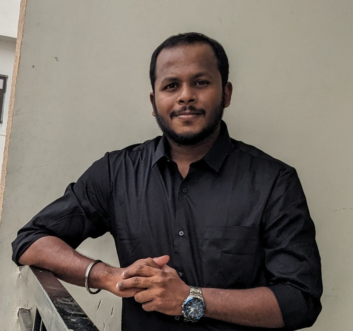
Mohamed Nijas
Research Area: Optical Coherence Tomography, Optical Fiber probes, Tumour Imaging, Lensless Microscope, Optical System Design.
My main research is to develop an optical fiber-based Spectral-domain OCT system. The system is primarily used for clinical validation of Gastrointestinal tumours. Also, I focus on the design,
fabrication and testing of various optical fiber probes for OCT. One such application is the Optical guidance system for epidural space identification. This system provides visual feedback to the
medical practitioner during the exploration of epidural space identification during epidural injections.
Awards
1.Best paper award in International conference on optics and Optoelectronics ICO-2019, IRDE, Dehradun, 19-22 October 2019
2. Best poster Award in Optical Society of India International Symposium on Optics, 20-22 IIT Kanpur, September 2018.
Email id: bm17resch11004@iith.ac.in
Quick Links: Google Scholar |
ResearchGate|
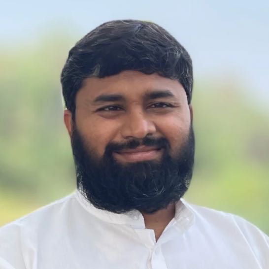
Dr. Prashanth Panta
Research Area: oral cancer, optical coherence tomography .
My research is focused on the use of intensity data in optical coherence tomography (OCT) for the classification of oral mucosal tissues. Our machine learning model identifies oral cancer with high accuracy. We have also conducted pilot studies in oral cytology, based on buccal smears. .
Samsung Innovation Award (2018) for "a smartphone based method to study cell for early detection of oral cancer"
Email id: bm18resch11006@iith.ac.in
Quick Links: Google Scholar |
ResearchGate |
LinkedIn |

Vikas Thapa
Research Area: Quantitative phase imaging (QPI), Generative Adverserial Network (GAN) for phase imaging.
Utilize capability of GAN for retrieving phase information of biological samples with single shot recording. This will enable high throughput, and compact QPI microscope.
Email id: bm18resch11017@iith.ac.in
Quick Links: Google Scholar |
ResearchGate|
LinkedIn |
GitHub |
Homepage
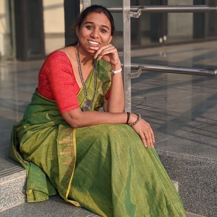
Vidya Gopal
Research Area: Biomedical signal processing, Machine learning and Deep learning.
My research centers on biomedical signal processing, leveraging machine learning and deep learning to advance vascular health monitoring. I am particularly passionate about developing cutting-edge deep learning applications for assessing cardiovascular and carotid artery health, aiming to improve early detection, risk stratification, and clinical decision-making through innovative computational approaches.
Email id: bm20resch11021@iith.ac.in
Quick Links: LinkedIn |
Google Scholar |
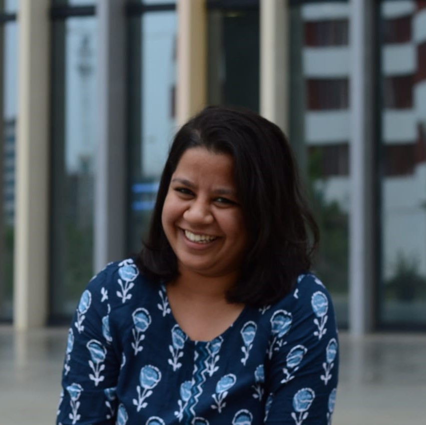
Greeshma Nechikat
Research Area: Microfabrication, Microfluidics, Lab-on-chip, point of care device, Electrochemistry, Organ-on-chip.
The aim is to develop biosensors for monitoring cancer prognosis post-chemotherapy and to develop and characterize Organ-on-Chip device for in-vitro applications.
Email id: bm21resch11007@iith.ac.in
Quick Links: LinkedIn |
Google Scholar |
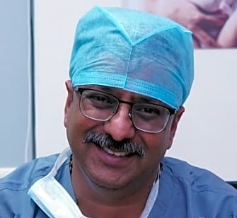
Dr. Sandeep Karunakaran
Research Area: In vitro fertilization (IVF), optics, microscopy.
The aim of my research is to improve the key performance indicators (KPIs) of In vitro fertilization (IVF) using better optics.
Email id: bm21resch14002@iith.ac.in
Quick Links: LinkedIn
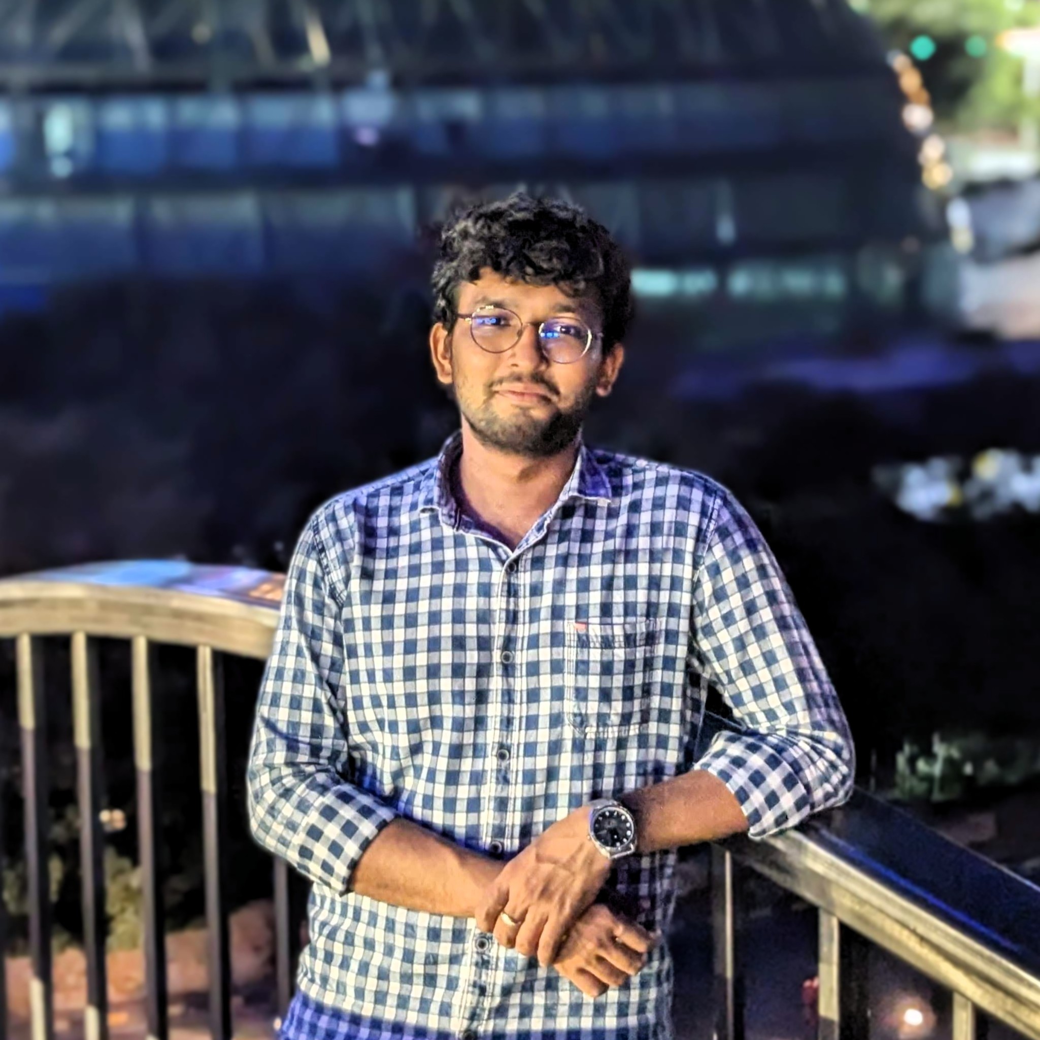
Mridul Verma
Research Area: Photoacoustic, Ultrasound, Signal & Image Processing, Beamforming Algorithms, Biomedical Optics, BM Imaging.
We work on developing Photoacoustic Imaging setup (PAM, PAT). Here, we work on optical systems, raw photoacoustic signal acquisition, signal processing, beamforming algorithms, and image processing. Tissue-mimicking phantoms are fabricated to simulate the properties of human tissue in order to test the imaging setup. We also work on the computational aspects, including MATLAB k-wave toolbox, NIRFast, and Monte Carlo Simulations.
Email id: bm22resch11004@iith.ac.in
Quick Links: LinkedIn |
Google scholar |
GitHub |
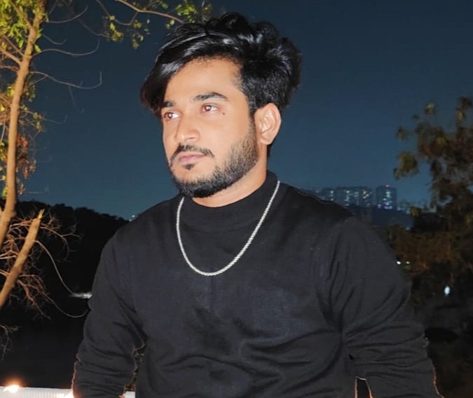
Sameer Mirza
Research Area: Microstructure property relationship in biological fluids.
Developing unique microrheological techniques to elucidate microstructure-viscoelastic property relationships for biological fluids. Optical, magnetic and acoustic probes will be leveraged to test
extremely small volumes of biological fluids which is expected to lay the groundwork to develop cheap in-situ biomedical tests for the future.
Email id: id22resch11005@iith.ac.in
Quick Links: LinkedIn |
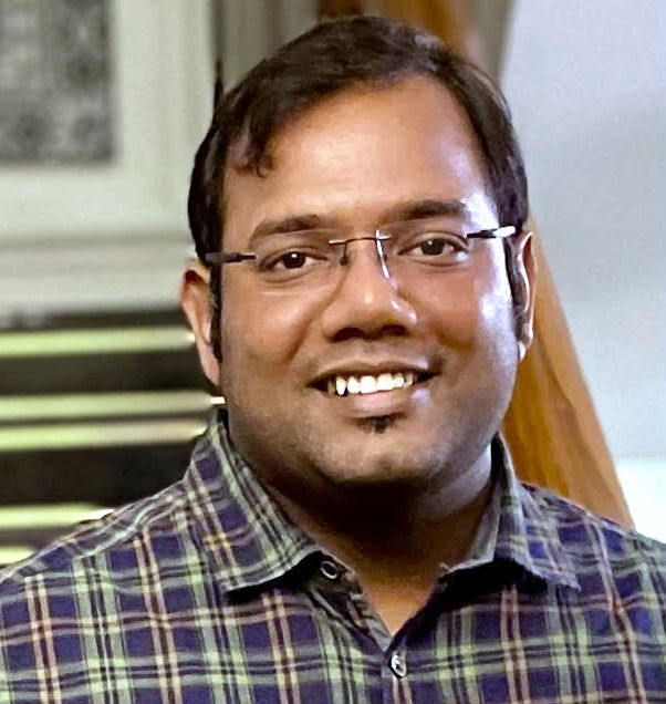
Saransh Khandelwal
Research Area: Biomedical Instrumentation, Non-Invasive Health Monitoring.
While being a part of the CSIR family, I worked in the core research involved in the Non-Invasive Estimation of Continuous Blood Pressure
Monitoring using Acoustic Sensors at CSIR-CSIO, Chandigarh as well as on the contrary gained experience in the field of Procurement and High-End Installation and
Commissioning of Modular OTs, MGPS, CSSD, Brachytherapy, LINAC and various other medical equipment for prestigious medical Institutes such
as AIIMS-Patna/Gorakhpur & IMS-BHU under PMSSY Phase II & III by MoHFW while working at a govt. PSU i.e. HLL-HITES, Noida . Currently, I am involved in both Academic and Research areas and working as
Senior Technical Superintendent at the Department of Biomedical Engineering, IIT Hyderabad.
Email id: bm23resch04003@iith.ac.in
Quick Links: LinkedIn
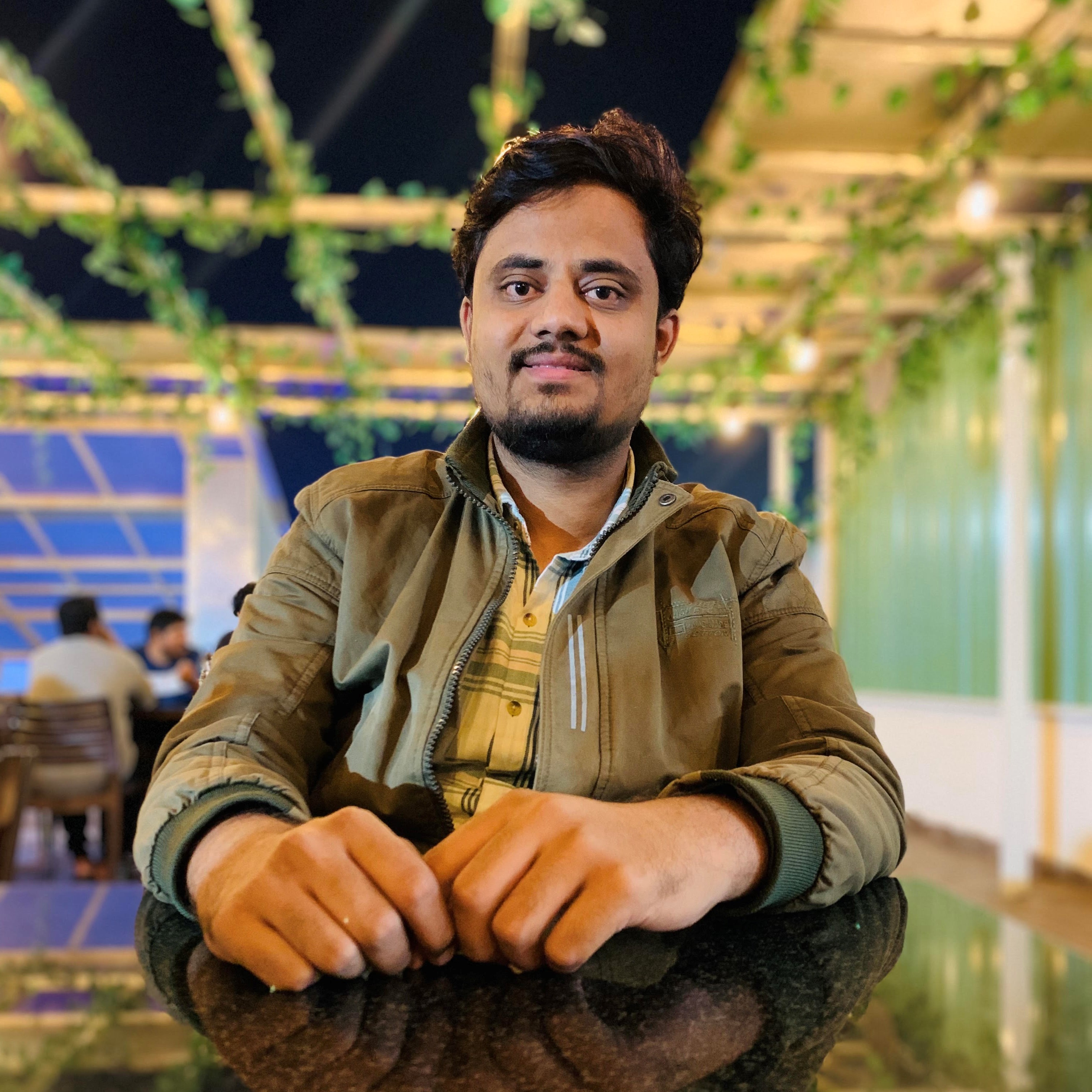
Sarfaraj Mirza
Research Area: Image Reconstruction, Deep Learning, GAN, Super Resolution Microscopy.
My research delves into the exciting frontier of medical optics: Physics-Informed Deep Learning for Super Resolution in Structured Illumination Microscopy. This approach aims to break the optical diffraction limit and offers two-fold resolution over conventional wide-field microscopes, enabling us to visualize subcellular biological structures. Master’s in Biomedical Instrumentation and over three years of research experience in image processing, prototype designing, deep learning, and signal processing at esteemed institutions, including CSIR-CSIO Chandigarh, PGIMER Chandigarh, and TIH-IIT Bombay.
Email id:bm23resch11008@iith.ac.in
Quick Links: Google Scholar |
LinkedIn|
ORCID |
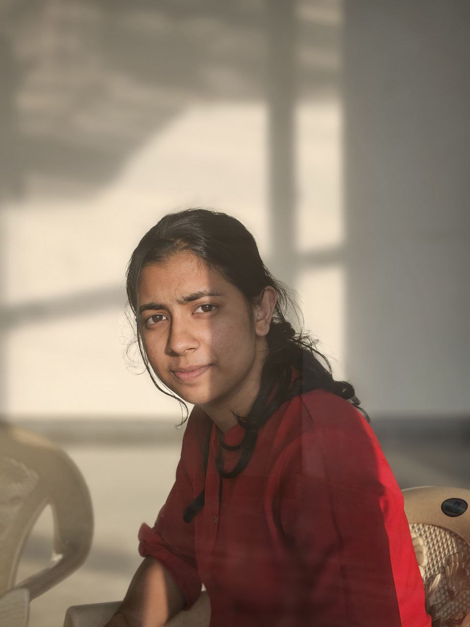
Jyothika
Research Area: Quantitative Phase Imaging (QPI), White Light Diffraction Phase Microscopy (wDPM).
I have completed my M.Sc. in Physics and M.Tech. in Optoelectronics and Laser Technology from Cochin University of Science and Technology (CUSAT). My research experience includes working on White Light Diffraction Phase Microscopy, with a keen interest in Quantitative Phase Imaging. Throughout my academic journey, I have focused on developing advanced imaging techniques and exploring their applications in biomedical optics.
Email id: bm24resch11010@iith.ac.in
Quick Links: LinkedIn
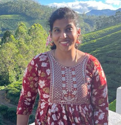
Sowmiya Sunil
Research Area: Tissue engineering, and biomedical diagnostics,.
Technologist in Biology. I am a researcher specializing in the development of organ-on-chip platforms for disease modelling and drug toxicity testing. My work lies at the intersection of
microfluidics, tissue engineering, and biomedical diagnostics, with a focus on creating physiologically relevant in vitro models that mimic the complex microenvironment of human organs. Also
interested to explore multi-organ integration on chip, while incorporating of biosensing elements for real-time monitoring of cellular responses.
Email id: sowmiya.sunil@bme.iith.ac.in
Quick Links: LinkedIn
MTech Students
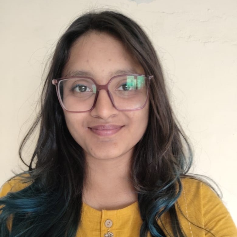
Devanshi Soni
Research Area: Optical coherence tomography (OCT), Speckle Imaging.
Analysis of scattering phenomena in Optical Coherence Tomography (OCT) images to improve image interpretation and diagnostic precision.
Email id: op24mtech11004@iith.ac.in
Quick Links: LinkedIn

Pratyushee Ghosh
Research Area: Optical Coherence Tomography (OCT), 3D Retinal Image Segmentation, Medical Imaging AI, Biomedical Instrumentation
Working on the development of an in-house OCT spectrometer system for biomedical imaging and implementing AI-based 3D segmentation of retinal layers to enhance diagnostic applications. Focus areas include hardware integration, image reconstruction, and deep learning models for ophthalmic disease detection
Email id: bm24mtech14005@iith.ac.in
Quick Links: LinkedIn
Our Alumni
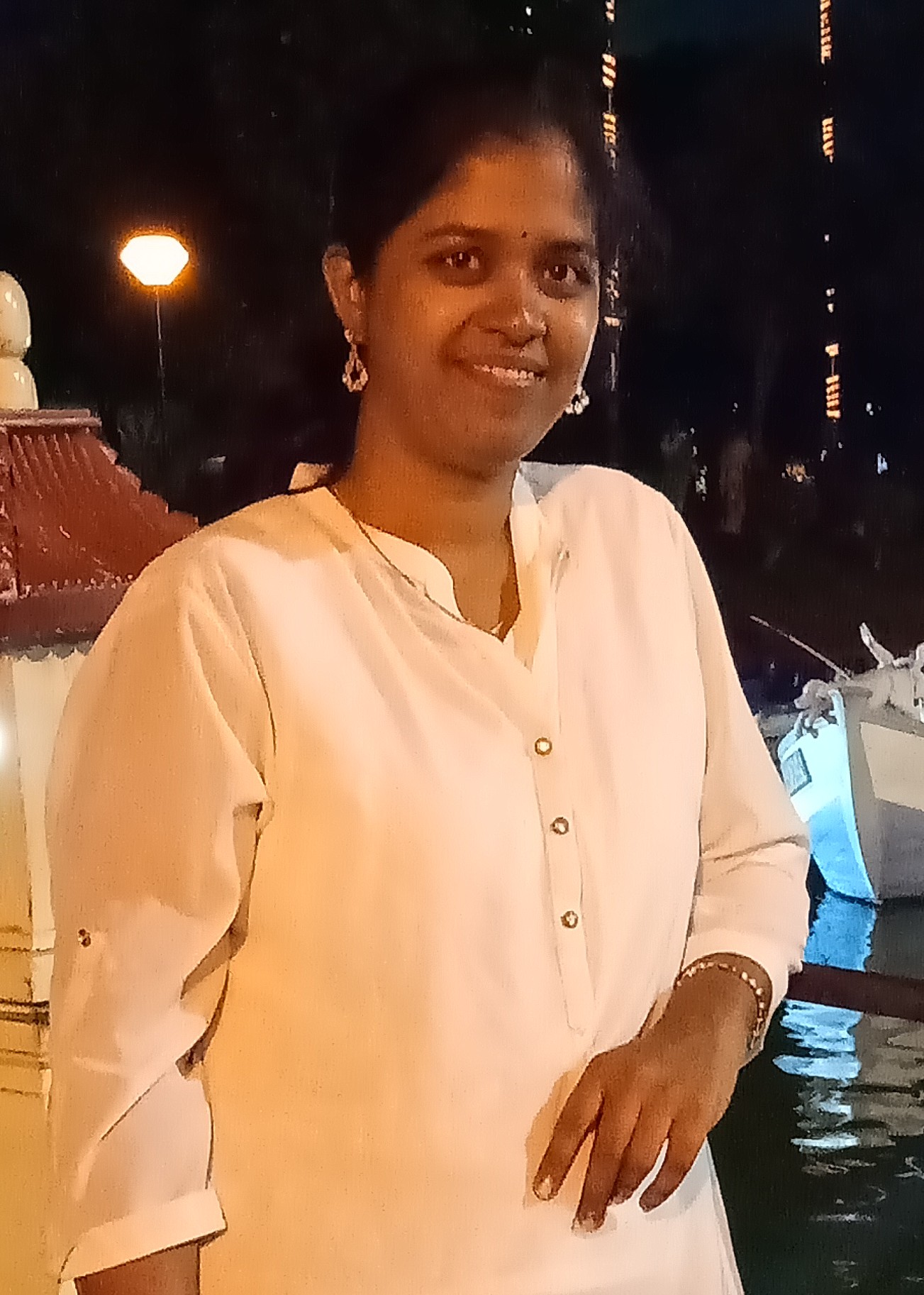
Ashwini S. Galande
Research Area: Lensless Holographic Microscopy for Point-of-Care Applications
A two-fold solution for the problems with conventional cytology procedures used for cervical cancer diagnosis. First is to develop
a low-cost, portable lensless holographic microscope, which gives quantitative phase information to screen cervical
cells, over the bulky and costly optical microscopes. The novel contribution here is the proposed sparsity assisted
phase retrieval algorithm to remove twin images and noise from the reconstructed image in real-time. Second is the
Deep Learning-based automated analysis to assist the doctor in diagnosis, so that there will be low false-negative
results, over the time-consuming manual examination of cell samples that results in inter-observer variability.
Email id: bm18resch11014@iith.ac.in
Quick Links: Google Scholar |
ResearchGate
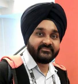
Amandeep Singh
Research Area: Optical Coherence Elasto-graphy imaging for biological samples using Digital holography Microscopy and related imaging techniques. optimization in phase reconstruction methods.
Being CSIR-UGC Fellow, I am pursuing my PhD from Bio-medical Optics lab collaborated with Bio-fab Lab. I have completed my Master's from Department of Physics, Punjab University, Chandigarh. As JRF fellow, I gained research experience from Medical Physics Lab (IISER Mohali) and currently working on development of imaging system for quantitative measurement of Elastography properties of biological samples.
Email id: bm18resch11016@iith.ac.in
Quick Links: Google Scholar |
ResearchGate|
LinkedIn |
Homepage

Dr.Inayathullah Ghori (Cardiologist)
Research Area: Low cost innovation to fight COVID crisis (Urja Project).
Application of AI/ML (Artificial Intelligence) algorithms for cardiac imaging.
Understanding the limitations of Clinical Decision Support Systems.
Hardware for tele-medicine projects. Participation & enabling of colleague's disruptive projects (Lab on a chip,
Phase Imaging). Design effective clinical training programs and making clinical pathways.
Email id: bm14resch11001@iith.ac.in
Quick Links: ResearchGate

Pawan Kumar
Research Area: Full-Field Optical Coherence Tomography (FF-OCT)
Optical Coherence Tomography is a low coherence interferometric imaging technique that is capable of
providing high resolution cross-sectional images of thick specimens. Full-Field Optical Coherence
Tomography (FF-OCT) is a variant of OCT that creates a transverse cross-sectional or en-face image at a
depth of a biological sample without doing any lateral scan. It is technically versatile for many clinical as
well as industrial applications.
Email id: bm16resch11003@iith.ac.in
Quick Links: Google Scholar |
ResearchGate
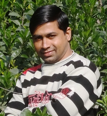
Nawab singh
Research Area: Micro/Nanofabrication, Nanomaterials Synthesis, Microfluidics, Point-of-Care Diagnostics, Lab-on-a-Chip, Nano-Bio Interface, Electrochemistry, Functional group chemistry, Biomolecules (Protein Biomarkers, Enzymes, Cancer Cell line, DNA), and Bio-conjugation, Surface Plasmon Resonance, Optical/Electrochemical Biosensor.
Email id: bm14resch11002@iith.ac.in
Quick Links: Google Scholar |
ResearchGate
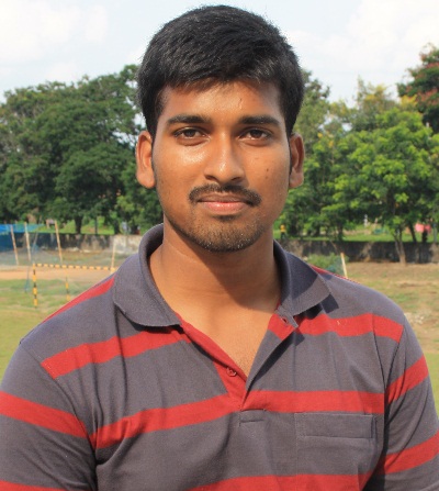
G Hanu Phani Ram
Research Area: quantitative phase imaging, multiple beam interferometry, microscopy. Digital Holography,
Inline-lensless holography, Oral cancer detection
Samsung Innovation Award (2018) for "a smartphone based method to study cell for early detection of oral cancer"
Email id: bm12m14p000001@iith.ac.in
Quick Links: Semantic Scholar |
ResearchGate
Shiv Kumar R
Research Area: Study and investigation of algorithms for the computer-aided diagnosis of retinopathy of prematurity in retinal fundus images of preterm infants.
Retinopathy of prematurity (ROP) is a sight threatening disorder that primarily affects preterm infants. It is the major cause for lifelong vision impairment and childhood blindness. Digital fundus images of preterm infants
obtained from a Retcam Ophthalmic Imaging Device are typically used for ROP screening.
ROP is often accompanied by Plus disease that is characterized by high levels of arteriolar tortuosity and v
enous dilation. The recent diagnostic procedures view the prevalence of Plus disease as a factor of prognostic
significance in determining its stage, progress and severity.
Our aim is to develop a diagnostic method, which can distinguish images of retinas with ROP from healthy ones and that can be interpreted by medical experts.
Email id: bm14resch11006@iith.ac.in
Quick Links: Google Scholar
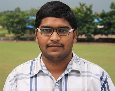
Praveen Kumar P (PhD)
Research Area: Non-interferometric quantitative phase imaging.
His work involved development and characterization ofTIE based quantitative phase microscope. Characterization of depth information of human sperm cell was his significant work.
Email id: BM13P1003@iith.ac.in
Quick Links: Google Scholar
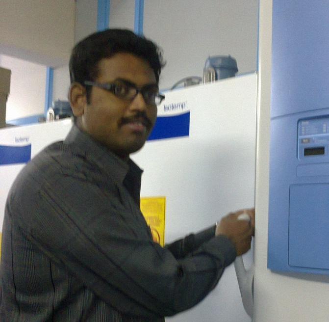
P Vimal Prabhu (PhD)
Research Area: Digital Holographic Microscopy for live cell imaging.
His work involved development and characterization of off-axis and on-axis holographic microscopy platforms.
Imaging and optimizing phase retrieval algorithm of nearly on-axis setup.
Email id: BO11P1005@iith.ac.in
Quick Links: Google Scholar
M D Azhar Ali (PhD)
Research Area: Biosensors and Surface Plasmonics.
Microfluidic‐integrated biosensors: Prospects for point‐of‐care diagnostics. Microfluidics
involves the science and technology of manipulation of fluids at the micro- to nano-liter level. It is
predicted that combining biosensors with microfluidic chips will yield enhanced analytical capa-
bility, and widen the possibilities for applications in clinical diagnostics.
Email id: BO11P1002@iith.ac.in
Quick Links: Google Scholar
MTech Alumni
Prashant Kumar Jaiswal
Research Area:An effort to automate the process of retinoscopy by employing a system of HD camera attached to a mirror retinoscope setup combined with video processing pipeline
An Optometry graduate and Ophthalmic Engineering post-graduate, core interest in providing engineering solutions for ophthalmic problems through design & development of ophthalmic equipment & devices, med-tech innovation, and research & development, aimed at solving problems in Ophthalmic care.
Email id: op22mtech14003@iith.ac.in
Quick Links: LinkedIn
Arjun Bhattacharya
Research Area: Elastography, Biomedical Optics, Medical Instrumentation, AR/VR platform, Digital Simulation
Quantification of Tissue Elastography via Optical Imaging Methods Utilising diverse algorithms, simulation tools, and multimodal imaging methods to quantitatively examine the biomechanical characteristics of several phantoms, tissues and samples using elastography techniques.
Email id: bm23mtech14001@iith.ac.in
Quick Links: LinkedIn