CoE - ICMR
Medical Device and Diagnostics Mission Secretariats
ICMR has sanctioned a Centre of Excellence to IITH to foster innovation and product development in the field of Medical devices and Diagnostics. The funding is Rs. 15.2 Cr. The team is led by Prof. Renu John and other co-investigators. Click here for more details.
Coherence imaging optics (Multidisciplinary)
Spectral Domain Optical Coherence Tomogrampy (SD-OCT)
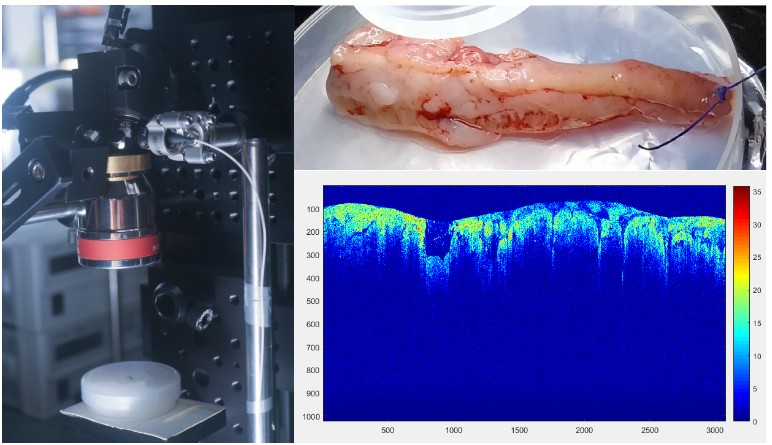
Optical Coherence Tomography (OCT) employs near-infrared light to illuminate biological tissues and gathers refracted/reflected light axially parcelled by coherence gating to produce high resolution, volumetric imaging. It holds great potential for early detection of superficial cancers and is therefore an essential diagnostic modality with ability to re-direct therapeutic interventions in the near future. We are working on Optical fibre-based OCT Probe for Clinical Imaging and tissue characterization.
Full-Field Optical Coherence Tomogrampy (FF-OCT)
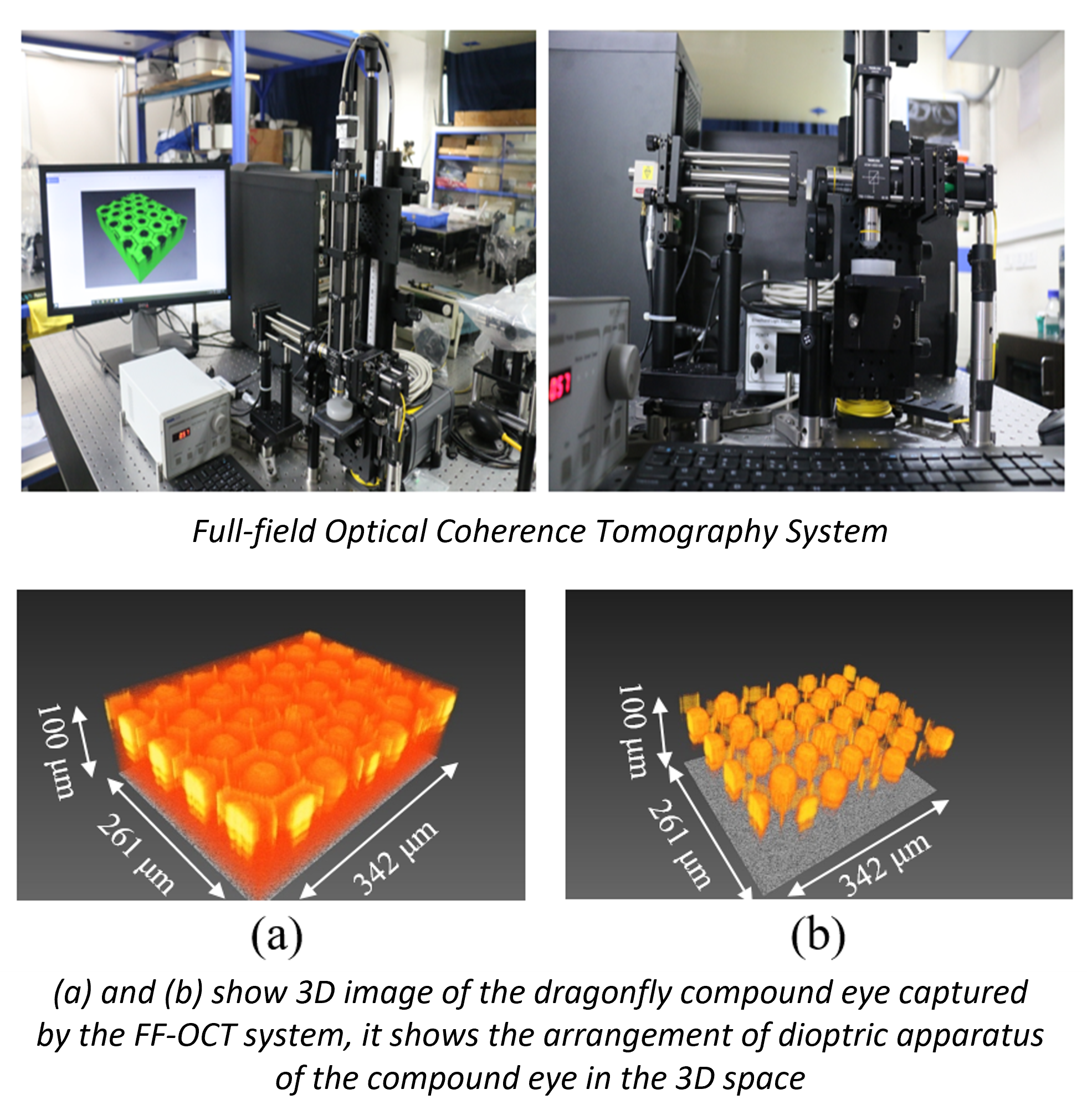
FF-OCT is a modified optical arrangement of the Optical Coherence Tomography System in which the whole field of view on the sample is illuminated simultaneously rather than point-by-point scanning, and the backscattered light from each point is detected in parallel. The parallel detection scheme improves the imaging speed, and the absence of lateral point scanning arrangements makes this system compact and mechanically less complex. The FF-OCT system images all the scatterers present at a fixed depth of the sample simultaneously and thus produces an en face cross-sectional image of the sample. Therefore, it is possible to use large numerical aperture microscope objectives with FF-OCT set-up to get higher lateral resolution. Thus, the FF-OCT approach overcomes the issues with traditional OCT systems due to the constraint between lateral resolution and depth of field. The lateral resolution of a FF-OCT system is controlled by the NA of the objective lens, while the bandwidth of the light source governs the axial resolution.
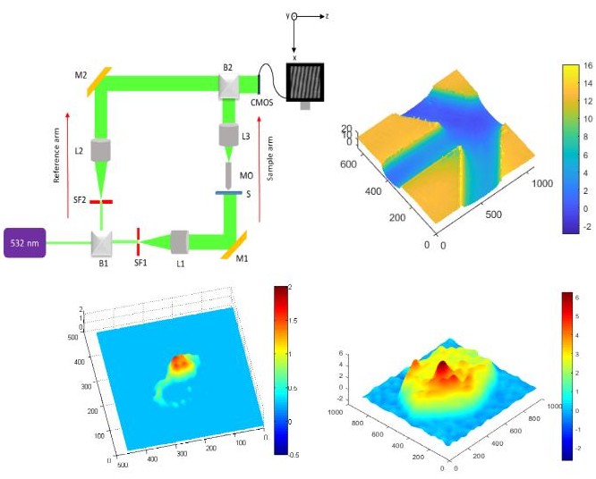
Digital Holography
Light microscopy is a key tool in clinical routine for observing biological samples. Tapping the phase information is indeed a vital step for label-free and non-destructive imaging. Quantitative phase imaging (QPI) would quantify the phase values and provide additional parameter in the form of thickness, thus volumetric imaging is made possible. The process of holography consists of two steps 1. Recording a hologram and 2. Object retrieval from recorded hologram. Invention of digital recording devices like CCD’s led to digital holography where object complex information is retrieved from numerical calculations using diffraction theory. Thus, numerical reconstruction is one significant aspects of digital holography. The zeroth order and twin image elimination in digital off axis holography can be carried out numerically. The propagation of the digital hologram to the object plane can be executed numerically using Fresnel Kirchhoff integral. We are focusing on imaging of unstained biological samples, their reconstuction and processing the phase images.
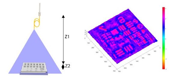
Lensless Inline Holography
The cost of the device plays an important role when it comes to the low resource settings. Integration of low cost devices with technology and big data analytics would play a major role in improving healthcare in developing countries. We aim to improve the quality of the retrieved phase and amplitude with minimal hardware and computational requirements for the lensless setup, thereby helping in the realization of a portable quantitative phase microscope that can be utilized in a variety of point-of-care healthcare applications. We are working on an inverse problem approach in the hologram plane, where the object information is searched that will best model the experimental hologram.
Optical Elastography
This project is highly focused on the assessment of mechanical stiffness for investigating the biomechanical properties of biological tissues. The Optical elastographic platform has been developed for the quantification of elastic along with viscous parameters for tissue characterization. First, the proof of concept studies has been conducted on tissue-mimicking phantoms of varying biomechanical properties and then, the same study has been performed on biological samples e.g. ex-vivo caprine liver, ex-vivo chicken liver, ex-vivo caprine kidney for qualitative along with a quantitative estimation of mechanical properties. The current studies are promising towards a non-invasive or minimally invasive imaging for in-vivo and ex-vivo mechanical characterization of tissues with future clinical applications.
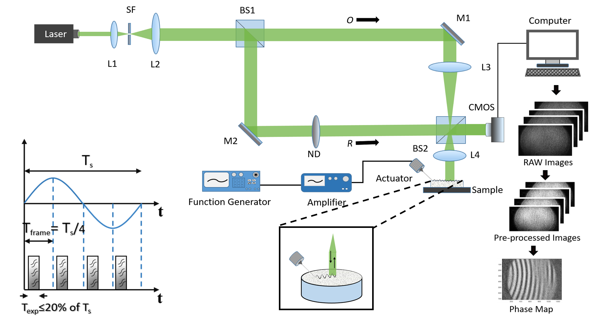
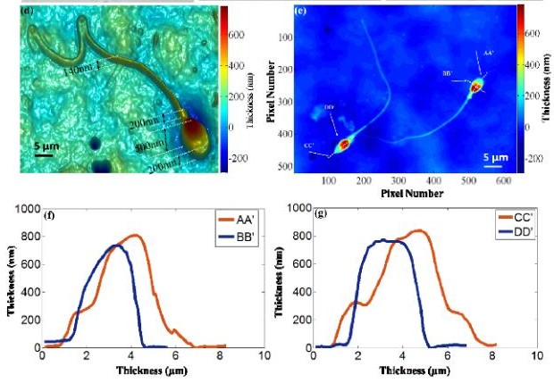
Transport of Intensity Equation (TIE)
Transport of Intensity Equation (TIE) is a non-interferometric quantitative phase imaging technique that is well-suited for this application due to its simplicity in experimental set-up and minimal computational requirements. TIE can be easily realized on a regular brightfield microscope with white light illumination. The phase image reconstructions of the object under study is possible using TIE by recording two defocus images and one in-focus image along the optical axis without the use of any phase unwrapping algorithms [32]. By exploiting a non-coherent white light source or a partially coherent light source [33–37], TIE is capable of reconstructing the thickness of the sample with nanometric depth precisions and high signal to noise ratio (SNR).
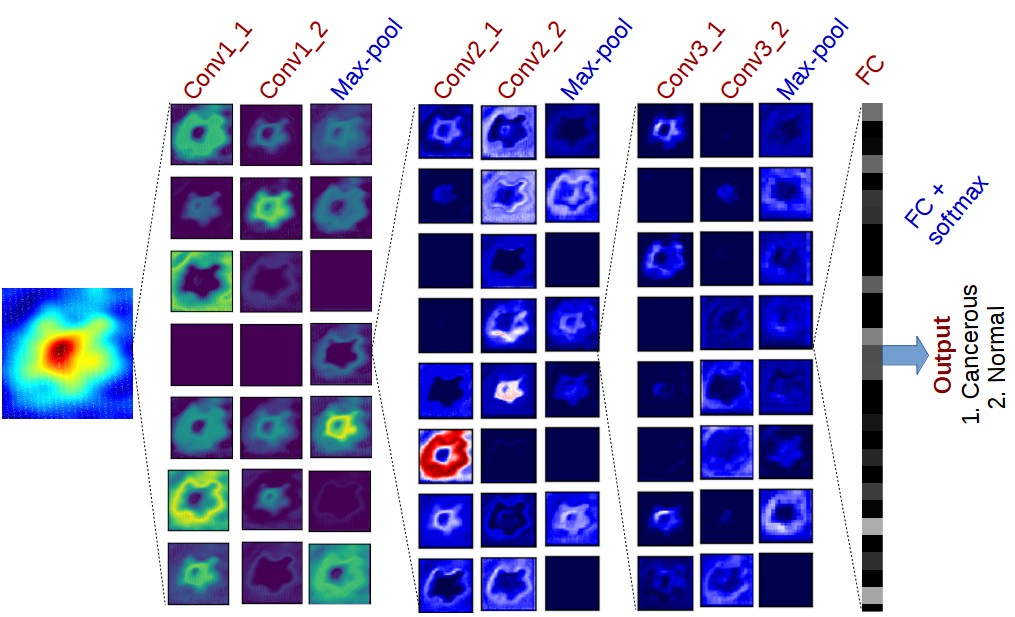
Learning Microscopy
Advancements in AI led the researchers to explore its utilization in quantitative phase imaging (QPI). The rapid label-free property of QPI enables it for the fast screening process for many biomedical imaging applications. Due to which QPI is the most preferable choice for AI assisted techniques. AI methods such as Deep learning (DL) are data-driver techniques and best suited to process a large amount of data. DL plays a vital role in terms of fast and early detection of various diseases by utilizing QPI data. DL is vital in terms of fast and early detection of many diseases. Currently we are working on building lensless microscope for cervical cancer detection using deep learning capabilities
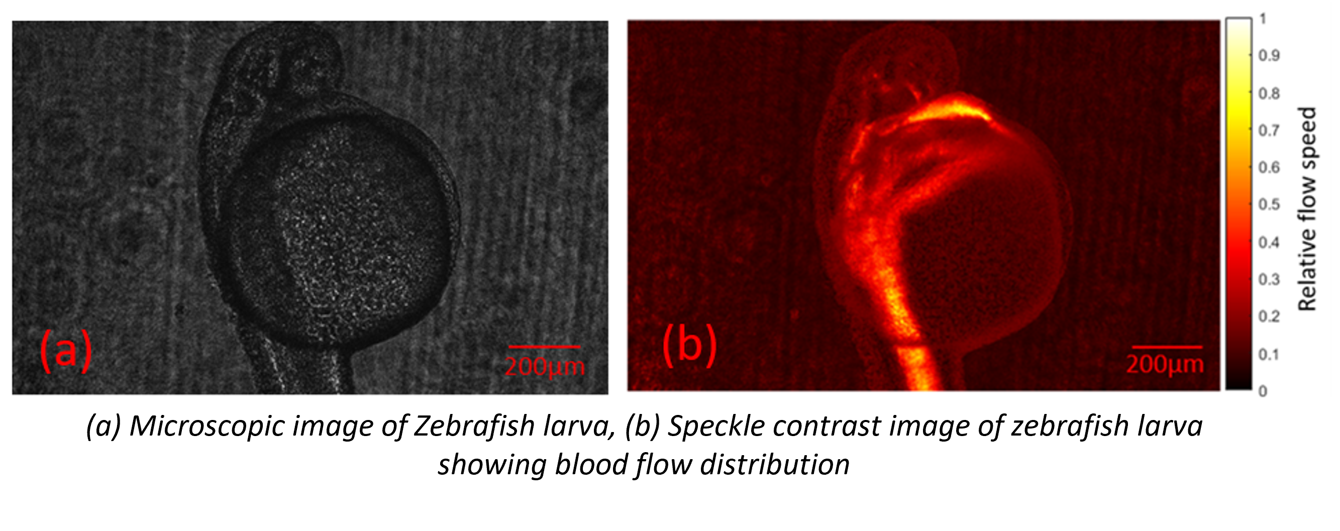
Speckle Contrast Imaging
Laser speckle contrast imaging is an imaging technique based on the principle of speckle formations. When laser light falls on optically rough surface the backscattered light will interfere and form a random interference pattern called speckle. Spatial and temporal variations of speckle pattern can quantify the motion of particles.
Nanomaterials Based Microfluidic Biosensors
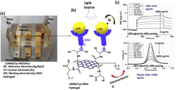
Dual-Modality Microfluidic Biosensor Based on Nanoengineered Mesoporous Graphene Hydrogels
Schematic representation of an incorporated dual-modality Cys-RGO hydrogel microfluidic chip for the detection of cMb. (a) A photograph of the nanoengineered integrated dual-modality mesoporous Cys-RGO hydrogel microfluidic biosensor chip. (b) Antigens-antibodies immunocomplex formation (c) Incorporated dual-modality Cys-RGO hydrogel sensor action revealing coupling of light and voltage source to measure electrochemical and SPR signals.
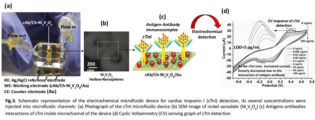
A hollow-nanosphere-based microfluidic biosensors for biomonitoring of cardiac troponin I
We were developed a microfluidic immunosensor using chitosan-nickel vanadate (Ch-Ni3V2O8) hollow-nanosphere for the detection of cTnI in human blood serum samples using electrochemical technique. This device offered a high sensitivity of 38.88 µA/ng mL‒1/cm2 with for a wide range of cTnI concentration (0.005‒100 ng mL‒1). We obtained a low limit-of-detection (LOD) of 5 pg mL‒1 of cTnI in human blood serum sample.
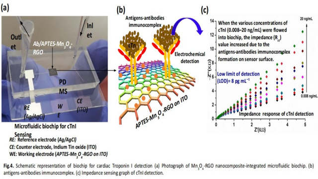
Microporous Nanocomposite Enabled Microfluidic Biochip for Cardiac Biomarker Detection
We were fabricated a microfluidic biochip using manganese-reduced graphene oxide (Mn3O4-RGO) nanocomposite for cTnI detection in serum samples using electrochemical Impedance technique. This device offered a high sensitivity of 87.58 kΩ/(ng mL−1)/cm2 for a wide range of cTnI concentration (0.008–20 ng/mL) with LOD of 8 pg mL‒1 of cTnI in serum with high stability, high reproducibility and good selectivity.
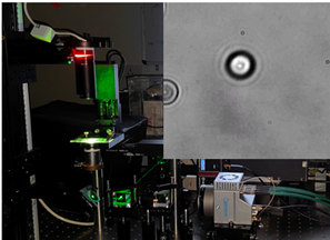
Optical tweezer
Optical tweezer is a minimally invasive, contactless and highly sensitive technique for 3D control over micro and nano length scale using a tightly focused laser beam. The manipulation capabilities of optical tweezers have laid the foundation for numerous applications in the fields of biological and physical sciences, understanding cellular processes, developing new therapies, and improving diagnostics. We aim to improve the landscape of clinical biomicrorheology utilising orbital angular momentum (OAM) and spin angular momentum (SAM) of light. We are working on interdisciplinary aspects of biology and physics to leverage the 3D micromanipulation for better understanding of fundamental yet pivotal physiological processes.